 |
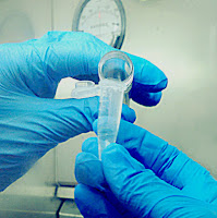
| The swab that used to collect specimen for influenza testing. |
Working in a Medical Lab
Great learning experiences in a Medical Laboratory.
Tuesday, January 5, 2016
Sunday, December 20, 2015
Various Cultures
Urine Culture
(Culture on Blood Agar and MacConkey agar)
(Culture on Blood Agar and MacConkey agar)
This test is performed when a patient is suspected to have UTI ( Urinary tract infection )
Basically, when there is growth of pure culture in high colony counts and the urinalysis resulted in large amount of bacteria, we will consider it as positive result for urine culture.
Methodology:
- Use a smaller loop to obtain the urine sample
- Draw a vertical line at first in order to distribute the sample evenly.
- Then streak the plate starting with close distance to far distance between each horizontal line.
 |
Blood Culture(Culture on Blood Agar and MacConkey agar)
This test is performed to determine and identify the presence of fungi, bacteria or virus in the sample. It is ordered when the patient shows the symptom of sepsis.
*Normally, the FBC( Full Blood Count ) test will also be ordered along to determine whether the patient have increased WBC (White blood cell ) that indicating a potential infection.*
1.
 |
| Scan the bar code. |
 |
| Insert the culture bottle into the appropriate position. |
3.
 |
| Wait 2 days for the analyzer to detect whether there is any bacteria growth inside the culture bottle. |
*Basically, there should be no bacteria inside the human blood as it should be sterile inside the body.*
4. If there is bacteria inside the culture bottle, a syringe will used to withdraw the patient's blood from the rubber sealed bottle and one drop of blood is used for culture.
 |
 |
Lancefield Grouping test :
-- a test that used to identify the species of detected Streptococcus.
 |
| Reagent used. |
 |
| There are total 6 types of Streptococus : A, B, C, D, F, G . The agglutination indicate that which the detected species belongs to. |
Swab Culture
(Culture on Blood Agar and MacConkey agar)
- If the culture consists of bacteria growth followed by beta-haemolysis, the microorganism is suspected to be the Streptococcus aureus.

β-hemolysis is the lysis of red blood cell in the blood agar around the colonies due to the streptolysin which produced by the bacteria. - Catalase test will be conducted.

Small amount of the suspected organism will mix with a drop of catalase which placed on a slide. Bubble formation indicating positive result. - Then, Coagulase test will be carry out.
Small amount of the suspected organism will mix with the plasma (FFP) within a test tube.
If there is coagulation of the plasma indicating positive result. - Gram staining and sensitivity test will take place afterward.
Stool Culture(Culture on MacConkey agar and XLD agar)
- Small amount of the patient's stool is added into a test tube containing Selenite F solution and incubate for 1 day.
- While in the same first day, the stool is also used to culture on MacConkey agar.
- In the second day, the incubated Selenite-F solution is cultured on XLD agar plate.
Positive result: Black colonies ( Salmonella ) present on XLD agar.
* The microbe will change the color of the agar*
Culture the "Black colonies" on TSI agar ( Triple Sugar Iron agar ).
(Left) Agar turned black indicating positive result with the presence of Salmonella.
(Right) Agar remained yellow in color indicating negative result.
Procedure of culturing on TSI:
- Stab vertically into the slant agar.
- Then, drag the loop out and streak a vertical line on the agar surface.
- Lastly, do the horizontal streaking.
Other culture
Upper respiratory sample( Sputum , throat swab...) ,Eye swab , high vagina swab... use Blood agar, MacConkey agar and Chocolate agar.
 |
| Chocolate agar is placed in a sealed tin with lighted candle while incubating in order to create an anaerobic environment. |
Other than Blood agar and MacConkey agar, urethral swab is also cultured on the Sabaraud agar to find out any yeast presented.
AFB testing
AFB testing ( Acid-Fast Bacillus Smear and Culture and Sensitivity )
- It is a test that used to diagnose tuberculosis ( caused be Mycobacterium tuberculosis) in the patient and help in monitoring the effectiveness of treatment.
- AFB test may also be used to detect any infection caused by the Mycobacterium species (Acid fast bacilli).
 |
| The pink rod-shaped microorganism is Mycobacterium tuberculosis |
Procedure:
- Prepare a smear and fix.

- Flood the smear with ZN-carbol fuchsin stain for 5 minutes and gently heat it.
- Wash gently with running tap water.
- Decolorize with TB-decoloriser for 2~3minutes
- Wash gently with running tap water.
- Counter stain with methylene blue for 30~60 seconds.
- Wash gently with running tap water.
- Dry over gentle heat.
- Examine under 100x with immersion oil.
Important notes during streak plate.
Important Notes
- Don't touch the wall using loop while streaking because the side wall of the agar plate is consider not sterile. ( except for Mueller Histon agar )

- Blood Agar (BA) should be streaked first before MacConkey (MAC) agar to avoid contamination by the bile salt and crystal violet in MAC which used to inhibit the growth of Gram-positive organism.
(Every microorganism can grow on BA)
(Left) MAC agar ; (Right) BA - Function of every types of used agar should be studied in order to determine which types of organism can be cultured on it.
- Sometimes when Urine FEME showed 3+ positive in the amount of bacteria, however there is no growth on the agar culture. This is because the bacteria observed under the microscope might be already dead or it could also be possible that there is mistake in the technique used to culture the plate.
- Gram-positive and gram-negative organism should not present together in certain part of the body. If the culture shows this type of result, we must always correlate with the FBC ( Full Blood Count ) result to see whether there is any infection.
C & S test
Culture and Sensitivity test
- The specimen is cultured on agar plate to identify the microorganism that present in the sample based on the culture's morphology, biochemistry profile and staining.
- The specimen is cultured on a specific antibiotic-containing agar plate to discover the microorganism based on its susceptibility.
Also, it helps in determining what type of antibiotic can be given to the patient for infection treatment. - This test can be done with different types of body fluids and excretion, such as urine, blood, pus, swab, saliva, mucus, stool and so on.
Procedure:
Culture and species determination :
Antibiotic Susceptibility Test:
Culture and species determination :
- Insert 4 full amount of normal saline into the test tube ( "Culture tube" )
- Take little amount of bacteria from the pure culture and insert into the tube until the saline looks slightly turbid.

- Incubate at 37°C for 2 hours.
- Insert 100µl of the culture into each well of the Microgen kit ( contains certain addictive and reagent for growing ) to determine the species of the microorganism.
- Add 2 drops of mineral solution.
- Seal the kit and incubate for 1 day.
- Add appropriate drops of reagent in to specific well.
- Compare the resulted color with the identification system chart, calculate the number and key in to the computer software to determine the species automatically.

Microgen kit which added with sample and reagent in each well.
 |
| Identification system for Streptococci and Enterococci . |
 |
| Identification system for all Enterobacteriaceae and excessive range of oxidase-positive Gram-negative bacilli. |
 |
| Fill in the blank and calculate the number. |
 |
| Key in details into the computer software. |
Antibiotic Susceptibility Test:
- Add 0.5ml of peptone water into a microcentrifuge tube ("Sensitivity tube" )


- Take little amount of bacteria from the pure culture and insert into the tube until the peptone water looks slightly turbid.

- Incubate at 37°C for 2 hours.
- Dip a clean cotton swab into the sensitivity tube and fully spread the solution on the agar plate ( Müeller-Hinton agar ).
- Add on the antibiotic chips with the appropriate distance ( about 2.4 cm) based on the species.
For example, Escherichia coli
For E.coli, there are 12 antibiotic chips to be tested. - Incubate the agar plate for 1 day.
Result Interpretation:
*Often correlate with other relevant test result before the final result is reported*
For example,
FBC result :
FBC result :
- High lymphocytes indicate viral infection, thus no bacteria will grow on the culture plate.
- High neutrophils indicate bacteria infection, thus there should be bacteria growth on the agar.
Reference:
Wednesday, December 16, 2015
WWF
 |
| 6 types of reagent . |
Procedure:
- Add 8000μl of saline in a container.
- Withdraw 100μl of the saline and replace it with the same amount of patient's serum.
- Add 1000μl of the diluted sample into 6 tests tubes which containing 6 different types of reagents in it. (1 drop in each)
- Incubate in water bath for 5 hours.
Result Interpretation:
- Positive result will shown in the sedimentation of the colored reagent.
Monday, December 14, 2015
Gram staining.
- Gram stain is used to differentiate the microorganism whether it is gram-positive or gram negative.

- Gram positive -Purple
Gram negative- Pink - It is conducted when the urine or swab culture have mixed growth of bacteria.
- For urine or stool culture, it is normal to have both gram-positive and gram-negative bacteria present in the culture.
- Except for stool, if there are more than 10 colonies appeared in other types of culture on blood agar, gram staining will be carry out for species identification.
Procedure:
 |
| Add caption |
- Prepare a smear and fix it by passing through gentle heating.
- Flood the smear with crystal violet for 1 minute.
- Rinse with running tap water.
- Flood the slide with iodine for 1 minute.
- Rinse with running tap water.
- Flood with decolorizer for 30 seconds.
- Rinse with running tap water.
- Counter stain the slide with safranin for 1 minute.
- Rinse with tap water and dry the slide.
- Observe under 100x with immersion oil.
Reference:
- http://www.medicinehack.com/2012/02/gram-staining-procedure-mechanism.html
- Labtestsonline.com
Subscribe to:
Comments (Atom)


