Wednesday, November 25, 2015

Reticulocyte Count

Procedure:

  1. 3 drops of blood + 1 drop of Brillant Cresyl Blue , and incubate for 15 minutes.
  2. After incubation, use the solution to make one thin blood film and view under microscope.


( count 1000 RBC )


[ Eg: If 1 field has roughly 100 RBC means have to look at 10 different fields. ]


Sunday, November 22, 2015

Blood Smear


Blood smear/ Blood film.
How to do a Blood Smear?

  1. Place a small drop of blood at one end of the smear to be made on the slide.
  2. Position the spreader (we use cover slip here) at 45° angle and backing into the drop of blood.


Differentiation of gender based on Blood Smear.

Female and male  have same amount of neutrophils bearing appendages. ( A to D form )

However, female has more A-form (drumstick) appendages while male has more C-form (tag) appendages due to higher androgen level in men.


References:

  1. http://www.ncbi.nlm.nih.gov/pmc/articles/PMC3781649/

Leishman stain


Thin blood smear.


Leishman stain
  • Acid-base staining
    • Acid (Red)    - bind to cytoplasm
    • Base (Blue)   - bind to nucleic acid (RNA, DNA...)


Procedures:
  1. Make a thin blood film by positioning the cover slip 30º~45º on a drop of blood, and wait it to dry completely.
  2. Then, place the slide on the rack and fully cover it with the Leishman stain.


  3. Wait for 4 minutes and add few drops of buffer with pH 6.5


  4. Gently blow or rock the slide to evenly mix the buffer with the stain.
  5. Wait for 12 minutes before washing those stains off from the slide with smooth running water.
  6. Allow the slide to be air dried.
  7. Look under microscope.

Examples:
The platelets are loosely distributed among the red blood cells.


Saturday, November 21, 2015

STart analyser / PT, aPPT & INR

Specimen: Patient's blood within the Sodium Citrate tube ( Blue top )


What is INR?
INR is known as the international normalized ratio.
It is used to monitor patient who has been treated with an anticoagulant, warfarin for blood thinning purpose.
It is a calculation for the appropriate drug dosage for patient based on the result of PT.


\text{INR}= \left(\frac{\text{PT}_\text{test}}{\text{PT}_\text{normal}}\right)^\text{ISI}


*normal=control
*ISI = ISI value for the analytical system being used.


Definition for PT test?
PT test is often used along with APTT to investigate on any bleeding or clotting disorder.

The PT evaluates the coagulation factor VII, X, V, II and I ( Fibrinogen ).
The APTT evaluates coagulation factor XII, XI, IX, VIII, X, V, II (Prothrombin), and I (Fibrinogen) , as well as PK (Prekallikrein) and HK (High Molecular Weigh Kininogen).


STart analyzer

*The blue pointer show the pipette tips*
*The red pointer shows the finnpipette*


Steel ball inside the cuvette ( pointed )


Methodology:
PT

  1. 50µl Plasma is inserted into a cuvette which containing a steel ball, and placed on the channel of the STart analyser.
  2. Next, start the timer and incubate for 60sec.
  3. After incubation, put the measuring test tube on the rack for the addition of reagent.
  4. 100µl of Neoplastine CI plus can be released from the finnpipette only when the release button is pressed.


APTT

  1. 50µl Plasma + 50µl CK Prest are inserted into a cuvette which containing a steel ball, and placed on the channel of the STart analyser.
  2. Next, start the timer and incubate for 180sec.
  3. After incubation, put the measuring test tube on the rack for the addition of reagent.
  4. 50µl of CaCl2 (0.025 pre-warmed) reagent is added.


Thursday, November 19, 2015

Difference between Plasma & Serum. ( & Anticoagulant )


Plasma consists of fibrinogen/clotting factor. It is the cells-free part of the blood which can be collected by spinning the red capped test tube that containing the blood until the blood cells have sediment at the bottom. The blood is often treated with anticoagulant before centrifuge.

Serum is the plasma without the clotting factors.
It can be obtained by centrifuge the blood sample after the blood is clotted in the SST tube.


Anticoagulant

  • Function: Block the formation of fibrin clot.
  • Examples:
    • EDTA
    • Potassium Oxalate
    • Sodium Citrate
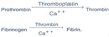
  • Mechanism: Removal of Ca++ which is essential for the conversion of prothrombin to thrombin.                       [Thrombin is the enzyme that initiate the conversion of fibrinogen to fibrin] 


Type of containers using in the Medical Lab.


Different tubes have different purposes and different additives.

Colour
Additive
Purpose
 Lavender Cap
 EDTA
 Anticoagulant
 Yellow Cap
 SST
 Separating Gel and Clot activator
 Red Cap
 Plain tube

 Sky blue Cap
 Sodium Citrate
 Whole blood

(Serum of patient which used for therapeutic drug monitoring should not be collected using serum separating tube. Instead, the specimen should be collected using a plain tube which containing no anticoagulant or additive. This is because the gel used for separating the serum will also isolate out  the lipophilic substances such as drug and produce a false result with low drug concentration.)

Yellow cap


Purple cap
Mini tubes for children.


Sky blue cap


Red cap
Mini tubes for children.


Swab -- For Influenza testing


http://www.questdiagnostics.com/home/physicians/testing-services/specialists/hospitals-lab-staff/specimen-handling/serum-plasma-whole-blood.html


Tuesday, November 10, 2015

Blood coagulation factors


Blood clotting factors
  1. Factor I          - Fibrinogen
  2. Factor II         - Prothrombin
  3. Factor III       - Thromboplastin
  4. Factor IV       - Calcium
  5. Factor V         - Proaccelein, Labile Factor
  6. Factor VI       - Accelerin
  7. Factor VII      - Proconvertin, stable factor
  8. Factor VIII     - Antihaemophilic globulin (A.H.G.)
                           - Antihaemophilic factor A (A.H.F.)
  9. Factor IX        - Christmas factor,
                            - Antihaemophilic factor B,
                            - Plasma thromboplastin component ( P.T.C.) 
  10. Factor X         - Stuart - power factor
  11. Factor XI        - Plasma thromboplastin antecedent ( P.T.A.)
  12. Factor XII       - Hageman factor
  13. Factor XIII      - Fibrin stabilizing factor


Coagulation factors are activated by:
  • Foreign surface
  • Tissue factor
  • Plasma protein factors
  • Calcium ions
  • Platelets


Classic blood coagulation theory of Morawitz

Haemostatic mechanism (Haemostasis) can be divided into 2 stages: Initiation stage and Maintenance stage. 


Initiation stage
Initiation factor - prevent bleeding from wound.
  • Temporary reflex nervous vasoconstriction of injured vessels.
  • Clumping of platelets to plug the injured vessels.
    Those platelets will release serotonin which resulted in constriction of blood vessel adjacent to the wound.
  • Tissue tension - The blood escape to the tissue space and causes swelling, thus increase the the tissue tension which further narrow down the surrounding blood vessel.

Maintenance stage
Formation of fibrin clot - involving many coagulation factors.